NEJM
Perspective
|
Teri Manolio, M.D., Ph.D.
A growing understanding of the importance of the rupture of atherosclerotic plaque in the pathogenesis of coronary events has led to the identification of an expanding array of markers of plaque vulnerability.1,2 Reports of markers of inflammation and coronary risk are appearing with increasing frequency; such markers include adhesion molecules, metalloproteinases, and as described in this issue of the Journal, myeloperoxidase (reported by Brennan et al., pages 1595�1604) and glutathione peroxidase 1 (reported by Blankenberg et al., pages 1605�1613). What should the clinician look for in these reports to determine their implications for patient care?
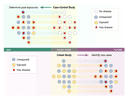
First, the measure should add independent information about the risk or prognosis. Risk can be assessed with the use of case�control and prospective cohort designs (see Figure), both of which are valid if they are carefully designed. These approaches produce estimates in the form of relative risks, odds ratios, or hazard ratios, comparing the incidence of disease among persons who have a given risk factor with the risk among those who do not have that risk factor. These measures should be adjusted mathematically for other factors that may account for some of the observed risk. Significant risk estimates, fully adjusted for other known confounders (not just age and sex), indicate that the marker adds independent information and thus has potential clinical importance. Applying this criterion to the study of myeloperoxidase reported by Brennan et al. and the study of glutathione peroxidase 1 reported by Blankenberg et al. shows that both markers provide independent information about risk.
|
Second, the measure should account for a large proportion of the risk associated with a given disease or condition. This is determined by the frequency of the risk factor and the magnitude of its associated risk. Hypertension, for example, is highly prevalent and increases the risk of congestive heart failure by a factor of 2 to 3, so that it may account for nearly half the cases of heart failure in a population.3 Typically, however, common risk factors do not have large relative risks (that is, they are not strong), and strong risk factors are not common, or nearly everyone would have the disease. When a high relative risk (more than 2 to 3) is first reported, one may wonder why it was not recognized previously or suspect that the estimate is biased. Risk can be overestimated by studying small, selected groups, using a control group that is at very low risk, or selecting extremes of a continuous measure for comparison (such as comparing the top with the bottom decile). The studies reported by Brennan et al. and Blankenberg et al. show that the risk and frequency of cardiovascular events associated with myeloperoxidase and glutathione peroxidase 1 are appreciable; both were associated with an increase in risk by a factor of 2 to 3 at levels present in 25 percent of the patients.
Third, the measure should be reproducible. A low coefficient of variation (standard deviation of repeated measures divided by their mean) is an indicator of good reproducibility; the coefficients of variation of glutathione peroxidase 1 were less than 10 percent in the study conducted by Blankenberg et al. The results should also not vary widely in an individual patient over time. Large variations in a measure that are unrelated to disease status, especially when their causes are unknown (as reported for some measures of hemostasis),4 reduce the practical value of a risk measure. Similarly, evidence of increased risk should be reproduced in multiple groups of patients and in a wide range of clinical settings; validation in several studies increases a clinician's confidence that the initial reports were not spurious. In this regard, myeloperoxidase levels have previously been associated with angiographically defined coronary disease5 and outcomes of acute coronary syndromes,6 whereas associations with glutathione peroxidase 1 activity have not been reported.
Fourth, if the measure is to be used as a diagnostic test, it should be sensitive and specific and have a high predictive value (see box). A highly sensitive test will be positive in nearly all patients with the disease, but it may also be positive in many patients without the disease. To be of clinical value, a test with high sensitivity should also have high specificity; in other words, most patients without the disease should have negative test results. For predicting the likelihood of disease on the basis of the test result, rather than the converse, the appropriate measures are positive and negative predictive values. Unfortunately, the positive predictive value falls as the prevalence of the disease falls, so tests for rare conditions will have many more false positive results than true positive results.
|
Most biologic markers, however, are not simply present or absent but have wide ranges of values that overlap in persons with a disease and in those without it. The risk typically increases progressively with increasing levels; few markers have a threshold at which the risk suddenly rises, so various cutoff points must be evaluated for their ability to detect disease. Cutoff points with high sensitivity, producing few false negative results, are used when the consequences of missing a potential case are severe, whereas highly specific cutoff points, producing few false positive results, are used to avoid mislabeling a person who is actually free of the disease. Sensitivity and specificity calculated at various cutoff points generate a receiver-operating-characteristic (ROC) curve, which ideally will be highly sensitive throughout the range of specificity. The most useful clinical tests are typically those with the largest area under the ROC curve, as Brennan and colleagues show for myeloperoxidase. The sensitivity, specificity, and predictive value of this measure, however, were only moderate, and in the case of glutathione peroxidase 1, Blankenberg et al. do not report these values.
Lastly, a test for the marker should be available. When first reported, many markers can be analyzed only in research laboratories. Results should be available rapidly enough to influence clinical decision making. Low cost and ease of performance are other desirable characteristics that are often strongly related to the availability of a test. Tests are not yet clinically available for either myeloperoxidase or glutathione peroxidase 1.
Initial reports of novel risk markers provide exciting clues to the pathophysiology of diseases, improved diagnostic capabilities, and ultimately, better patient care. Crossing the boundary from research to clinical application, however, requires replication in multiple settings, experimental evidence supporting a pathophysiologic role, and ideally, intervention trials demonstrating that modification improves the outcome. Until these steps are taken, specific interventions to modify novel markers should be reserved for investigative purposes. In the interim, however, these markers may be quite useful in identifying patients who may benefit from other therapies proven to reduce risk, since the risk generally increases with the number of risk factors present. Simple but often underused interventions, including blood-pressure control (achieved in only 30 percent of U.S. patients with hypertension), lipid-lowering therapy, smoking cessation, antiplatelet therapy, and after a myocardial infarction, use of beta-blockers and angiotensin-converting�enzyme inhibitors, may be particularly helpful in patients with other, as yet unmodifiable, coronary risk factors. Until their true implications for patient care are known, novel risk markers are thus best used to identify high-risk patients who would benefit from aggressive management of established risk factors.
I am indebted to Drs. Curt Furberg, Claude Lenfant, Eugene
Passamani, and Douglas Rosing for their critical review and comments.
Source Information
From the National Heart, Lung, and Blood Institute, National Institutes of Health, Bethesda, Md.
References
- Morrow DA, Braunwald E. Future of biomarkers in acute coronary
syndromes: moving toward a multimarker strategy. Circulation
2003;108:250-252.
[Full Text] - Pearson TA, Mensah GA, Alexander RW, et al. Markers of
inflammation and cardiovascular disease: application to clinical and public
health practice: a statement for healthcare professionals from the Centers
for Disease Control and Prevention and the American Heart Association.
Circulation 2003;107:499-511.
[Full Text] - Levy D, Larson MG, Vasan RS, Kannel WB, Ho KK. The progression from hypertension to congestive heart failure. JAMA 1996;275:1557-1562. [Abstract]
- Sakkinen PA, Macy EM, Callas PW, et al. Analytical and biologic variability in measures of hemostasis, fibrinolysis, and inflammation: assessment and implications for epidemiology. Am J Epidemiol 1999;149:261-267. [Abstract]
- Zhang R, Brennan ML, Fu X, et al. Association between
myeloperoxidase levels and risk of coronary artery disease. JAMA
2001;286:2136-2142.
[Abstract/Full Text] - Baldus S, Heeschen C, Meinertz T, et al. Myeloperoxidase serum
levels predict risk in patients with acute coronary syndromes. Circulation
2003;108:1440-1445.
[Abstract/Full Text]