NEJM
|
Thomas S. Kupper, M.D.
In this issue of the Journal, two reports present data on the efficacy
of two new biologic drugs for psoriasis. One of these drugs,
etanercept, has been used extensively in rheumatology and targets the
pleiotropic inflammatory cytokine tumor necrosis factor  (TNF-). The other, efalizumab,
targets CD11a, or L,
one chain of L
(TNF-). The other, efalizumab,
targets CD11a, or L,
one chain of L  2
integrin, also known as leukocyte-function�associated antigen 1
(LFA-1); LFA-1 is important in the process by which T cells cross
blood-vessel walls, enter tissue, and are subsequently activated by
antigen. Increasingly, molecules that were once the exclusive domain
of immunologic scientists have entered mainstream medicine.
2
integrin, also known as leukocyte-function�associated antigen 1
(LFA-1); LFA-1 is important in the process by which T cells cross
blood-vessel walls, enter tissue, and are subsequently activated by
antigen. Increasingly, molecules that were once the exclusive domain
of immunologic scientists have entered mainstream medicine.
Etanercept consists of the p75 TNF-
receptor fused to an IgG construct and binds to both soluble TNF-
and TNF- on the cell surface,
thus inhibiting its binding to cellular signaling receptors. TNF-
is made by leukocytes, including a subgroup of T cells, but can also
be produced by cells that are not derived from the bone marrow and
that reside in tissues, including skin. Etanercept has demonstrated
efficacy in rheumatoid arthritis, inflammatory bowel disease, and
psoriatic arthritis, and there are anecdotal reports of its efficacy
in additional immune and inflammatory diseases. A study in this issue
of the Journal (pages 2014�2022) indicates that it is also
effective in psoriasis.
Efalizumab is a humanized monoclonal antibody against CD11a that blocks LFA-1 interactions with intercellular adhesion molecules 1 and 2 (ICAM-1 and ICAM-2, respectively). LFA-1 is expressed only by leukocytes, and although T cells are not unique in their expression of it, they depend on LFA-1 for both successful extravasation and antigen presentation. A study of the efficacy of efalizumab in psoriasis is also reported in this issue of the Journal (pages 2004�2013).
To understand how these biologic agents work in psoriasis, it is
useful to take a step back and consider the roles of LFA-1 and TNF-
in the unique immunologic features of skin, our most exposed
interface with the environment.1 Skin is endowed
with special features that protect it from injury or infection, and
a limited number of factors, including the cytokine TNF-,
transmit danger signals from injured tissue to the immune system. The
release of TNF- from cells in
the skin induces the production of other cytokines and chemokines and
modifies endothelial surfaces in cutaneous postcapillary venules,
facilitating the extravasation of leukocytes. These leukocytes exit
vessels and enter the dermis through a multistep process involving
several molecules, including LFA-1. Leukocytes are then attracted
along chemotactic gradients and can begin to mediate effector
functions, such as the killing of pathogenic bacteria or fungi. One
of the prominent effector molecules produced by these infiltrating
leukocytes is TNF-. Fundamentally,
this process is a form of immunosurveillance of body surfaces for
danger signals, a phylogenetically ancient process that is central to
innate immunity.
Adaptive immunosurveillance is the domain of T cells. Each T cell
has a different specificity for antigen conferred by its unique
T-cell receptor, and getting the right T cell to the right place at
the right time is a major logistic challenge for the immune system.
This puzzle is solved by the specific migration patterns of different
subgroups of T cells.1,2
Naive T cells shuttle between blood and lymph nodes � a process
that is dependent on LFA-1. Once in lymph nodes, these T cells mingle
with dendritic cells that have recently migrated through the
lymphatics from the peripheral tissue. These dendritic cells have
left the peripheral tissue because danger signals (such as TNF-)
induced their migration and maturation, and they are uniquely
powerful activators of T cells that bear the correct receptor for
antigens they have internalized. T-cell activation depends on the
clustering of critical ligand�receptor pairs in immune synapses
that are at the interface of the T cell and the antigen-presenting
cell.3 These include the T-cell receptor
interacting with antigen bound to major-histocompatibility-complex molecules
(antigen recognition), T-cell CD28 binding to CD80 and CD86 (costimulation),
and T-cell LFA-1 binding to ICAM-1 on the dendritic cell.
When they are thus activated, naive T cells divide and multiply, express new molecules on their surface, and are instructed to become effector memory T cells. This immunologic memory extends to the anatomical location, so that a T cell that is educated in a skin-draining lymph node will express molecules that facilitate its subsequent entry into skin, whereas education in a Peyer's patch leads to the expression of molecules on T cells that facilitate their ultimate entry into the lamina propria of the gut.2 Skin-homing T cells express cutaneous lymphocyte antigen (CLA), CC chemokine receptors 4 and 10, and LFA-1 and will interact preferentially, in a sequential, multistep fashion, with blood vessels in skin that express E-selectin and P-selectin, CC chemokine ligands such as CCL17, and ICAM-1. There is evidence of the constitutive recruitment of skin-homing memory T cells into normal skin through this process.1,4 These memory T cells reside in the skin for an indeterminate period, and if they are not activated by their antigen, they enter lymph nodes through the lymphatics and ultimately return to the blood.
The pathologic release of TNF-
and other cytokines strongly up-regulates the expression of
endothelial E-selectin and ICAM-1, as well as chemokines, on the
luminal aspect of the skin vessels. Using these newly expressed
molecules, circulating skin-homing memory T cells can more
efficiently enter the skin through this multistep process, again with
the use of interactions between LFA-1 and ICAM-1. Once in the dermis,
these T cells may encounter their antigen, appropriately presented by
a dendritic cell or other antigen-presenting cell. Their successful
activation requires the formation of another immunologic synapse,
mediated in part by LFA-1�ICAM-1 interactions.
The immunosurveillance of the skin by skin-homing memory T cells is
fundamental to adaptive immunosurveillance and represents a powerful
means of protection against environmental pathogens. This elegant
system is subverted in psoriasis, because the immune system appears
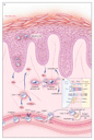
to perceive putative psoriatic autoantigens as foreign (see Figure).
In response to TNF-�mediated
danger signals, skin-homing memory T cells flood into the skin, and
those that are reactive to psoriatic autoantigens are activated, with
both processes involving LFA-1. Antigen presentation is also
facilitated by TNF- released by
non�T cells in skin, which leads to the maturation of dendritic
cells into more powerful antigen-presenting cells. The T cells that
are activated in the skin subsequently produce cytokines, and those
that are involved in psoriasis produce type 1 cytokines, including
interferon- and TNF-.
The release of TNF- by T cells
amplifies the inflammatory response, and in patients with a psoriatic
genotype, this leads to the characteristic hyperproliferative
response of the epidermis and the other cardinal features of
psoriasis. This process is self-perpetuating and is reversible only
when the activation of T cells within the lesions is blocked. Given
the activity of these molecules as described above, it is not
surprising that blocking either LFA-1 or TNF-
has therapeutic effects in psoriasis.
and TNF-.
The release of TNF- by T cells
amplifies the inflammatory response, and in patients with a psoriatic
genotype, this leads to the characteristic hyperproliferative
response of the epidermis and the other cardinal features of
psoriasis. This process is self-perpetuating and is reversible only
when the activation of T cells within the lesions is blocked. Given
the activity of these molecules as described above, it is not
surprising that blocking either LFA-1 or TNF-
has therapeutic effects in psoriasis.
|
Most therapies that are currently available for psoriasis have dose-limiting toxic effects, and the emergence of effective biologic agents that apparently have good safety profiles represents a triumph of translational research. These biologic agents, however, interfere with fundamentally important immunologic processes that have evolved over millions of years to protect the host from infection. Their use should not be undertaken lightly, since one would expect immunosurveillance to be altered, at least transiently, in persons who receive them. Certainly, this problem is not unique to biologic agents: cyclosporine, methotrexate, and other commonly used drugs come with similar caveats.
How does one evaluate claims that seem to favor one drug over another?
Proponents of alefacept, the first biologic agent approved by the
Food and Drug Administration for use in psoriasis, claim as one of
its advantages that it is memory�T-cell specific. The manufacturers
of efalizumab claim not only that it is T-cell specific, but that,
unlike alefacept, it does not reduce the numbers of T cells in
peripheral blood. The makers of etanercept claim that it, too, does
not reduce T-cell numbers and that it is designed to focus on the
central role of TNF- in psoriasis.
At this point, there are insufficient data to support claims that
one of these agents is superior to another. The relative importance
of these molecules and their targets can only be guessed at;
moreover, their activity may vary from person to person. It may be
that pharmacogenomics is an important variable: there may be groups
of people who have a better response to one or the other of these
agents, perhaps because of polymorphisms in genes that control the
expression of the relevant molecules. One thing is certain: we have
not seen the last of biologic therapies for psoriasis, and this will
ultimately be a boon to patients with this chronic, debilitating
disease.
Source Information
From the Department of Dermatology, Brigham and Women's Hospital, Boston.
References
- Robert C, Kupper TS. Inflammatory skin diseases, T cells, and
immune surveillence. N Engl J Med 1999;341:1817-1828.
[Full Text] - von Andrian UH, Mackay CR. T-cell function and migration: two
sides of the same coin. N Engl J Med 2000;343:1020-1034.
[Full Text] - Dustin ML, Colman DR. Neural and immunological synaptic
relations. Science 2002;298:785-789.
[Abstract/Full Text] - Kunkel EJ, Boisvert J, Murphy K, et al. Expression of the
chemokine receptors CCR4, CCR5, and CXCR3 by human tissue-infiltrating
lymphocytes. Am J Pathol 2002;160:347-355.
[Abstract/Full Text]