 UK
Europe
USA&Canada
Aust.&NZ
Asia
UK
Europe
USA&Canada
Aust.&NZ
Asia
Editorials Online�
Medical
Law News
E-texts
Links
Classifieds
Home
|
|
|
NEJM
Barry J. Maron, M.D.
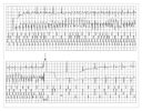
The time you won your town the race / We chaired you through the market-place; Man and boy stood cheering by, / And home we brought you shoulder-high. To-day, the road all runners come, / Shoulder-high we bring you home, And set you at your threshold down, / Townsman of a stiller town. � A.E. Housman, "To an Athlete Dying Young" (1895) Young athletes have come to be regarded as a special part of society, owing to their unique lifestyle and the widely held perception that they epitomize health and invulnerability, capable as they are of admirable and sometimes extraordinary physical achievements.1,2,3 Indeed, the possibility that young, highly trained high-school, college, or even professional athletes may harbor potentially lethal heart disease or be susceptible to sudden death under a variety of circumstances4,5,6,7,8,9,10,11,12,13,14,15,16,17,18,19,20,21,22,23,24,25,26 seems counterintuitive. Nevertheless, such sudden cardiac catastrophes continue to occur, usually in the absence of prior symptoms, and they have a considerable emotional and social impact on the lay public and physician community. Attempts to understand the causes of such events have triggered considerable interest in differentiating physiologic athlete's heart from structural cardiovascular disease,27 as well as in developing preparticipation screening strategies14 and formulating disqualification criteria15 aimed at preventing sudden death in young athletes. The Magnitude of the Problem The precise frequency with which sudden death occurs in young athletes (those under 35 years of age) remains unresolved. In Minnesota, the annual incidence of sudden death due to undiagnosed cardiovascular disease is reported to be about 1 in 200,000 high-school athletes participating in organized sports.16 However, such data are limited, and the magnitude of this public health problem may be considerably underestimated. Regardless of prevalence, when an athlete dies suddenly, the substantial social and emotional effect on the community is largely due to the youth, apparent good health, and lost potential of the athlete. Once regarded as personal and family tragedies, the unexpected death of an athlete now often becomes part of the public discourse and is fueled by media. Cardiovascular Causes of Sudden Death Although the overall population of athletes is at generally low risk for sudden death,5,16 a number of largely congenital but clinically unsuspected cardiovascular diseases have been causally linked to sudden death in young trained athletes, usually in association with physical exertion.3,4,5,6,7,8,9,10,11,12,17 In large autopsy-based surveys of populations of athletes in the United States, hypertrophic cardiomyopathy has consistently been the single most common cardiovascular cause of sudden death (Table 1), accounting for about one third of these events in prior reports.6 Hypertrophic cardiomyopathy, characterized by an asymmetrically hypertrophied and nondilated left ventricle, is a relatively common genetic cardiac disease (with an incidence of 1 in 500 persons in the general population),29 with heterogeneous clinical, morphologic, and genetic expression29,30,31 (Figure 1A). Sudden death is probably a consequence of an electrically unstable and unpredictable myocardial substrate with reentrant ventricular tachyarrhythmias (Figure 2), as evidenced by the presence of histopathological markers of disorganized myocardial architecture (Figure 1B) and replacement scarring (the consequence of microvascular abnormalities and myocardial ischemia).29,30
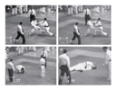
The second most frequent cardiovascular cause of sudden death on the athletic field is congenital coronary-artery anomalies in which the artery arises from the wrong aortic sinus (most commonly, the left main coronary artery originates from the right sinus of Valsalva) (Table 1).3,4,5,6,9,17,18,19,28,32 These anomalies, which are not usually associated with coronary-artery atherosclerosis, may be more common than previously thought.18,33 Their diagnosis requires a high index of suspicion and is particularly important, since surgical correction is feasible.17,33 The possibility of a coronary anomaly should always be considered in a young athlete with a history of chest pain or syncope, particularly if the episodes are triggered by exercise.34 Transthoracic or transesophageal echocardiography and magnetic resonance imaging can be used to diagnose the anomaly,17,19 and diagnostic coronary arteriography can ultimately be performed. Patients usually do not have abnormalities on 12-lead or exercise electrocardiograms,17,18,19 because the myocardial ischemia is episodic, thereby limiting the value of random screening. The most likely mechanisms of ischemia include acute-angled kinking at the origin of the coronary artery or compression of the anomalous artery between the aorta and pulmonary trunk during exercise. A diverse array of other, largely congenital malformations accounts for the remaining sudden deaths from cardiovascular disease among athletes3,4,5,6,7,8,9,10,11,12,13,16,17,18,19,20,32,33,35,36 (Table 1 and Figure 1). These include conditions known to cause sudden death, such as valvular heart disease (aortic stenosis or myxomatous mitral-valve degeneration), atherosclerotic coronary artery disease (Figure 1F), dilated cardiomyopathy (Figure 1D), Marfan's syndrome, arrhythmogenic right ventricular cardiomyopathy (Figure 1E), hypoplasia and other rare coronary anomalies, and myocarditis (Figure 1C). Each is responsible for only a small minority of the deaths (i.e., 5 percent or fewer)5,6 (Table 1). Myocarditis is challenging to diagnose clinically (or even at autopsy) and may be suggested in the absence of symptoms on the basis of its electrocardiographic abnormalities alone, which include heart block and ventricular arrhythmias.15,36 The diagnostic yield may be enhanced by using the reverse-transcriptase�polymerase-chain-reaction assay to identify a viral genome in endomyocardial-biopsy specimens.37 About 2 percent of young athletes who die suddenly have normal cardiac structure at autopsy, and no definitive cause of death can be established.3,5,6 Such deaths are probably due to conditions that are not associated with gross cardiac abnormalities � for example, ion-channel disorders (the long-QT syndrome38 and the Brugada syndrome39), the Wolff�Parkinson�White syndrome,40 structural abnormalities of the conducting system and microvasculature,12,41,42 catecholaminergic polymorphic tachycardia,43 right ventricular outflow tract tachycardia,44 coronary vasospasm, undetected segmental arrhythmogenic right ventricular cardiomyopathy,45 or subtle morphologic forms of hypertrophic cardiomyopathy.14,29,30 Intramural tunneled coronary arteries (short segments of the left anterior descending coronary artery that are surrounded by myocardium) are occasionally the sole abnormality found at autopsy.5,6,46 Such anomalies may have important clinical implications during intense athletic activity.46,47 In athletes with heart disease, primary ventricular tachyarrhythmias are the predominant mechanism of sudden death, although in athletes with Marfan's syndrome, death is often due to a ruptured aorta.6,35 The demographic profile of athletes who died suddenly in the Veneto region of northeastern Italy differs from that reported in the United States. In Veneto, arrhythmogenic right ventricular cardiomyopathy is reported to be the most common cause of death on the athletic field.7,10,13,48,49 This difference may be due to a particular genetic predisposition in the Italian population, or it may be a direct result of the unique national program of screening all young athletes before they can participate in organized sports.48 This program probably results in the identification and disqualification from competition of disproportionately fewer athletes with arrhythmogenic right ventricular cardiomyopathy than of those with more readily identifiable diseases (e.g., hypertrophic cardiomyopathy).3,14,45,48 Sudden death due to cardiovascular disease occurs in a wide variety of sports, but it has been reported most commonly among basketball and football players in the United States5,6 and among soccer players in Europe.7,10,13,45 Sudden death is much more common in male athletes (by a ratio of 9 to 1),5,6 probably because young women have lower rates of participation in certain sports (e.g., football).3,5 Hypertrophic cardiomyopathy is the most common cause of sudden death from cardiac causes in young black male athletes with previously undiagnosed cardiac abnormalities, a finding that contrasts sharply with the underrepresentation of blacks in clinically identified populations of patients with hypertrophic cardiomyopathy.28 This observation suggests that socioeconomic status and ethnic background may have major effects on access to diagnostic tests for hypertrophic cardiomyopathy.28 The association of unsuspected cardiovascular disease and sudden death in young athletes is not coincidental, since participation in competitive sports itself substantially increases the likelihood of sudden death.6,49 Furthermore, up to 90 percent of deaths among young athletes occur during training or competition.1,3,5,6,15,16,28,49 These observations substantiate the finding that in the presence of certain cardiovascular diseases, vigorous physical exertion represents a trigger for lethal arrhythmias and sudden death on the athletic field. Sudden death is not, however, limited to competitive athletes and may occur in nonathletic young persons during recreational or even sedentary activities.4,8,9,10,26,29 Athletic-Field Risks Unrelated to Cardiovascular Disease Sudden death may also occur under diverse circumstances among sports participants who do not have underlying heart disease.21,22,23,24,25,50,51,52,53 Commotio Cordis Most notable examples of sudden death in athletes without antecedent heart disease occur as a result of blunt, nonpenetrating, and innocent-appearing blows to the chest that produce ventricular fibrillation unassociated with structural injury to the ribs, sternum, or heart21,51 (Table 1 and Figure 3). The precise frequency of commotio cardis during athletic events is unknown, but it may be a more common cause of sudden death than many of the cardiovascular diseases known to cause these catastrophes (Table 1).
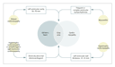
The precordial blows that trigger commotio cordis are often not perceived as unusual for the sporting event involved or of sufficient magnitude to cause death (Figure 3). Commotio cordis is most common in children and adolescents (mean age, 13 years), since these age groups characteristically have compliant chest walls that probably facilitate the transmission of the energy from the chest blow to the myocardium.21,51 Sudden death due to commotio cordis is often caused by being struck by sports projectiles, such as baseballs and hockey pucks. Such projectiles have a broad range of velocities but most commonly strike the precordium with only moderate force. Examples include a pitched baseball traveling at 30 to 40 miles per hour (48 to 64 km per hour) and a hockey puck or lacrosse ball traveling at 90 miles per hour (144 km per hour). Commotio cordis is also occasionally caused by physical contact such as a karate blow (Figure 3) or collisions between outfielders chasing a baseball.21,51 Many deaths from commotio cordis also occur during informal sporting activities among friends and relatives around the home or at the playground. They may also occur accidentally during everyday activities unrelated to organized sports, such as a chest blow delivered in an effort to relieve hiccups.51 Unfortunately, some of the latter deaths have triggered criminal convictions for manslaughter or murder.54 Survival after commotio cordis is uncommon (15 percent) and is most likely when cardiopulmonary resuscitation and defibrillation are prompt.21,51 Findings in a swine model that replicates commotio cordis have provided important insights into the mechanisms responsible for the devastating electrophysiological consequences of precordial blows.55,56,57,58 To provoke ventricular fibrillation, the blows must be directly over the heart56 and occur within 15 to 30 msec before the T-wave peak (which represents about 1 percent of the cardiac cycle) during the vulnerable phase of repolarization.55,56,57,58 Some spontaneously aborted episodes of commotio cordis may result from blows sustained during depolarization, since such activity triggered transient complete heart block in the swine model.55 Basic cellular mechanisms responsible for commotio cordis are incompletely understood, although selective activation of potassium-ATP channels may have a pivotal role.57 Several strategies for the prevention of commotio cordis, including innovations in the design of sports equipment, have been considered. Although the use of softer-than-normal ("safety") baseballs reduces the risk of ventricular fibrillation under laboratory conditions,55 it does not provide absolute protection against sudden death on the baseball field.51 Chest barriers with proven efficacy for youth sports (e.g., baseball, lacrosse, and hockey) are not yet commercially available, and some available products do not protect the precordium completely.21,51 Automatic external defibrillators save lives in the community because they can elicit a rapid response and early defibrillation and automatically provide an analysis of cardiac rhythm.59,60,61 If these systems become more widely disseminated and available for use by the general public at schools and athletic facilities, they will undoubtedly result in the survival of many athletes who have cardiac arrest as a result of blows to the chest or cardiovascular disease.62 Other Risks A small number of sudden deaths are reported each year among athletes that are due not to cardiac causes but to factors such as extreme heat, leading to hyperthermia and central nervous system dysfunction (heat stroke)5,23,52; head and spine trauma (usually among football players and pole vaulters)22; uncontrolled bronchial asthma; ruptured cerebral-artery aneurysm; sickle cell trait53; and nonpenetrating blows to the neck by hockey pucks, which trigger rupture of the vertebral artery and subarachnoid hemorrhage.63 Sudden unexpected death, nonfatal stroke, and acute myocardial infarction in trained athletes have been attributed to the abuse of cocaine, anabolic steroids, and dietary and nutritional supplements.24,25,50 Dietary supplements such as ma huang, an herbal source of ephedrine (i.e., elemental ephedra), which is a potentially arrhythmogenic cardiac stimulant,25,50 are often taken to enhance athletic performance or to mask the presence of other drugs during testing. Causal linkage between the use of dietary supplements and cardiovascular events is largely inferential, based on a close temporal relation between the ingestion of the compound and adverse events in otherwise healthy people. Athlete's Heart Systematic training in predominantly endurance sports (dynamic or aerobic) or isometric sports (static or power) triggers increases in cardiac mass and structural remodeling in many athletes.64,65,66,67,68,69,70,71,72,73,74,75,76,77,78,79,80,81,82 This physiologic form of hypertrophy, or athlete's heart, is regarded as a benign adaptation to systematic athletic training with no adverse cardiovascular consequences.64,65,66,67,68,69,70,71,72,73,74,75,76,77,78,79,80,81,82,83,84,85,86,87 The resultant changes include enlargement and increased volume of the ventricular chambers,65,68,72,74 sometimes accompanied by increased thickness of the left ventricular wall64 and an increase in the size of the left atrium, with preservation of systolic and diastolic function. The magnitude of the physiologic increases in cardiac mass vary according to the sport; extreme changes in cavity dimensions and wall thickness have been reported most commonly during training for rowing, cross-country skiing, cycling, and swimming64,65,72,80 but, paradoxically, less commonly during training for ultraendurance sports.67,68 Isometric training (e.g., weight lifting or wrestling) is associated with left ventricular wall thickness that is usually normal in absolute terms (less than 12 mm) but disproportionately increased in relation to cavity size.70,71,72,83 The angiotensin-converting�enzyme genotype has been associated with the magnitude of exercise-induced left ventricular hypertrophy in endurance athletes, suggesting that genetic factors may have a role in this process.88 The abnormal cardiac dimensions associated with athletic training are related to body-surface area or lean body mass64,65,66,69,70,71,72,75,76 and are consequently less pronounced in female athletes.66,75 Other physiologic adaptations to training include a variety of abnormal patterns on 12-lead electrocardiograms in about 40 percent of athletes,84 some of which resemble those of cardiac disease (greatly increased voltages, Q waves, and repolarization abnormalities).84,85,86 Owing to the heightened vagal tone that accompanies physical conditioning,89 trained athletes without structural heart disease are also subject to many arrhythmias and conduction alterations which usually do not require invasive investigation or specific treatment � premature atrial and ventricular beats, sinus tachycardia or bradyarrhythmia, supraventricular tachycardia, junctional rhythm, and first-degree, or Wenckebach, atrioventricular block (Mobitz type I). Frequent ventricular premature beats and complex ventricular ectopy, such as couplets and nonsustained ventricular tachycardia, may also be present on ambulatory Holter monitoring87 and may be difficult to distinguish from ectopy associated with inflammatory myocardial disease caused by viral agents (often enterovirus but also adenovirus),36,87 long-term abuse of cocaine,24 or vector-borne pathogens.90 Athlete's Heart and Cardiovascular Disease The ability to make clinical distinctions between physiologic athlete's heart and pathologic conditions27 has critical implications for trained athletes, since reducing the risk of sudden death or progression of cardiovascular disease may be the basis for the disqualification of athletes from competitive sports.15 Alternatively, overdiagnosis may lead to unnecessary restrictions, depriving athletes of the psychological or monetary benefits of sports. Indeed, the morphologic adaptations of athlete's heart may mimic cardiovascular disease and lead to a differential diagnosis that includes hypertrophic and dilated cardiomyopathy and arrhythmogenic right ventricular cardiomyopathy27,64,65,66,69,80,84 (Figure 1 and Figure 4). Such diagnostic dilemmas frequently arise when cardiac dimensions fall outside clinically accepted partition values (left ventricular wall thickness of 12 mm and cavity size of 60 mm). About 2 percent of highly trained adult male athletes have mild increases in left ventricular wall thickness (13 to 15 mm)64; female and adolescent athletes have somewhat lower cutoff values. Such changes fall into a gray area in which extreme expressions of athlete's heart and mild morphologic forms of hypertrophic cardiomyopathy overlap (Figure 4).27 This diagnostic ambiguity can often be resolved through the use of a number of noninvasive measurements, such as the response of cardiac mass to short periods (about three months) of deconditioning,77,79,80,82 or assessment of diastolic filling with Doppler echocardiography.27,81 DNA-based diagnostic tests that can definitively distinguish genetic heart diseases from athlete's heart are not yet available on a routine clinical basis.27,29
The extreme alterations in cardiac dimensions evident in some athletes have inevitably raised the question of whether such exercise-related adaptations are truly physiologic and benign. For example, about 15 percent of highly trained athletes have striking enlargement of the left ventricular cavity, with an end-diastolic dimension of 60 mm or more, similar to that occurring in dilated cardiomyopathy65 and difficult to distinguish from pathologic states, particularly when the ejection fraction is at the lower limit of normal. A longitudinal echocardiographic study showed incomplete reversal and substantial residual dilatation of the chambers in 20 percent of retired, deconditioned elite athletes.82 Although firm evidence is lacking, we cannot rule out the possibility that extreme ventricular remodeling associated with intense conditioning may have adverse clinical consequences over the long term.82,89 Preparticipation Screening One objective of systematic medical evaluations in large, general populations of trained athletes before competition is to detect "silent" cardiovascular abnormalities that could progress or cause sudden death.14 Indeed, the identification of asymptomatic patients with genetic diseases such as hypertrophic cardiomyopathy, arrhythmogenic right ventricular cardiomyopathy, the long-QT syndrome, and the Brugada syndrome (defined by right bundle-branch block and ST-segment elevation in the right precordial leads) has taken on even greater importance, since high-risk patients can have a cardioverter�defibrillator implanted for the primary prevention of sudden death (Figure 2).91,92,93 However, the presence of an implanted cardioverter�defibrillator is not a sufficient reason in and of itself to allow an athlete to return to competition.2 Major obstacles to the implementation of preparticipation screening are the large number of young athletes eligible for evaluation (about 8 million in the United States) and the rarity of sudden death from cardiovascular causes in this population (estimated overall prevalence, 0.5 percent or less).3 Customarily, screening of U.S. high-school and college athletes consists of history taking and physical examination,14,94,95,96 a strategy that lacks sufficient power to identify important cardiovascular abnormalities consistently. For example, although the nonobstructive form of hypertrophic cardiomyopathy is the single most common disease entity responsible for sudden death in young athletes, screening can be expected to raise the suspicion of or identify this disorder relatively infrequently, because potential diagnostic markers such as a loud heart murmur in a supine person at rest, syncope, and a family history of sudden death6,14 are usually absent. In a retrospective study, only 3 percent of trained athletes who died suddenly of heart disease had been suspected of having cardiovascular abnormalities on the basis of preparticipation screening involving history taking and physical examination, and none were disqualified from competition after screening.6 The quality of cardiovascular screening of U.S. high-school and college athletes has come under scrutiny.94,95,96 A major impediment lies in the design of (and guidelines for) approved questionnaires.94,95,96 The guidelines examiners are given for screening high-school athletes are inadequate in 40 percent of the states when measured against the recommendations of the American Heart Association (AHA). These include, for example, history of exertional chest pain or excessive dyspnea, family history of heart disease, or heart murmur.14 Furthermore, legislation in several states allows health care workers with vastly different levels of training and expertise (including chiropractors) to conduct preparticipation sports examinations, often under suboptimal conditions.14 Improvements in the screening process related to history taking and physical examination would undoubtedly result in the identification of greater numbers of athletes with previously undiagnosed but clinically relevant cardiovascular abnormalities. National standardization of high-school (and college) screening medical examinations, incorporating AHA recommendations,14 would be the most practical approach to achieving this goal. For more than 30 years the Italian government, as a result of the Medical Protection of Athletic Activities Act, has mandated national preparticipation screening and medical clearance of all young athletes who want to participate in organized sports programs.48 The annual evaluations routinely include history taking, physical examination, and a 12-lead electrocardiogram. Since the electrocardiogram is abnormal in up to 95 percent of patients with hypertrophic cardiomyopathy,29 this program permits the identification of many athletes with previously undiagnosed disease.48 Similar screening efforts in the United States, albeit in smaller populations, have had less productive results.97,98,99 Obstacles to implementing obligatory government-sponsored national screening in the United States involving electrocardiography, echocardiography, or both include the particularly large population of athletes, major cost�benefit considerations, and recognition that it is not possible to eliminate the risks associated with competitive sports.14,15 However, some volunteer-based screening programs have emerged, including ones in which portable echocardiographs are used to screen high-school athletes in the field.100 In large populations, however, the value of preparticipation screening with the use of noninvasive tests is limited by the expected large numbers of false positive (i.e., borderline) results, as well as false negative results in which subtle but important lesions go undetected � for example, when echocardiography is performed in the prehypertrophic phase of hypertrophic cardiomyopathy (in persons under 14 years of age)29 or when coronary anomalies are not recognized.17 The preferred diagnostic strategy for athletes who are initially suspected of having cardiovascular disease (e.g., because of findings on screening or the presence of symptoms) should focus on the systematic exclusion of structural heart diseases known to cause sudden death in young people, beginning with a history taking and physical examination, electrocardiography, and echocardiography. Syncope is a particularly challenging symptom in young athletes that requires careful investigation to resolve critical distinctions between physiologic events such as neurocardiogenic (neurally mediated) syncope and those related to underlying heart disease. Criteria for Sports Eligibility and Disqualification When a cardiovascular abnormality is identified in a competitive athlete, there are several questions to consider: What is the risk of sudden death if the athlete continues to participate in organized sports? Would the risk be reduced if the athlete stopped training and competing? Which criteria should be used to determine the athlete's eligibility for (or disqualification from) athletic competition? However, the disqualification process can become polarized, given the personal desires and aspirations of the athlete and the mandate of the physician to protect patients from circumstances that incur unacceptable risks.1,2,3 The recommendations of the 26th Bethesda Conference offer clear benchmarks for clinical practice.15 The guidelines for athletic eligibility or disqualification are predicated on the premise that intense training and competition increase the risk of sudden death in susceptible athletes with heart disease and that this risk is likely to be reduced by temporary or permanent withdrawal of the athletes from sports.2,6,14,15,28,48,49 Indeed, the unique pressures of organized sports do not allow athletes to exercise strict control over their level of exertion or reliably discern when cardiac symptoms arise that make it prudent to terminate physical activity. Under the current Bethesda guidelines, young athletes with unequivocal hypertrophic cardiomyopathy are discouraged from participating in competitive sports, with the exception of low-intensity sports such as golf and bowling.15 Some acquired diseases that may be reversible (such as myocarditis) justify temporary withdrawal of an athlete from competition, followed by resumption of organized sports activity if resolution is documented.15 A U.S. appellate court2 ruled that guidelines such as the Bethesda Conference report15 can be used by team physicians to formulate appropriate decisions about an athlete's eligibility and thus also set a precedent for resolving future medicolegal disputes involving college athletes. For sports participants with cardiovascular abnormalities, the risk associated with intense physical exertion cannot be quantified precisely, given the extreme and unpredictable physiological conditions to which they may be exposed. Indeed, not all sudden deaths from hypertrophic cardiomyopathy are associated with intense physical activity,29 and not all trained athletes with this disease die suddenly during competition.3,28 Although formally controlled studies are lacking, indirect evidence and clinical intuition suggest that screening and disqualification strategies are well justified and probably reduce the number of sudden deaths in young athletes.14,48,49 Decisions to remove high-profile athletes with cardiovascular disease from competition may be confounded by complex social ramifications and can prove difficult to implement, particularly when collegiate or professional careers are at stake.1,2,3 Many elite athletes with heart disease may not fully appreciate the implications of the medical information presented and are too often willing to accept risks and resist recommendations to stop competing in order to remain in the athletic arena.1,2 However, any waivers of responsibility that they may sign are not necessarily legally binding or enforceable.2,15 In these emotionally charged circumstances, physicians' medical judgments can be insidiously affected by pressures exerted by the athletes' family members, coaches, and school administrators, as well as by the considerable confusion created by athletes who solicit multiple medical opinions until they find one that endorses their continued participation in sports.1,2,3 A measure of responsibility for these complex situations may well be attributable to materialism and societal attitudes, which exaggerate the importance of organized sports.1,2,3
Dr. Maron reports having received grant support from
Medtronic.
From the Hypertrophic Cardiomyopathy Center, Minneapolis Heart Institute Foundation, Minneapolis. Address reprint requests to Dr. Maron at the Hypertrophic Cardiomyopathy Center, Minneapolis Heart Institute Foundation, 920 E. 28th St., Suite 60, Minneapolis, MN 55407, or at [email protected]. References
|