 UK
Europe
USA&Canada
Aust.&NZ
Asia
UK
Europe
USA&Canada
Aust.&NZ
Asia
Editorials Online�
Medical
Law News
E-texts
Links
Classifieds
Home
|
|
|
NEJM
Jean Marie Naeyaert, M.D., Ph.D., and Lieve Brochez, M.D., Ph.D.
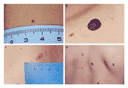
This Journal feature begins with a case vignette highlighting a common clinical problem. Evidence supporting various strategies is then presented, followed by a review of formal guidelines, when they exist. The article ends with the authors' clinical recommendations. A 26-year-old man comes to establish primary care. Physical examination reveals multiple moles on his body, which he describes as "funny-looking." There is no family history of melanoma. He thinks that one of his two brothers (15 years of age) and his father have the same kind of moles. How should this case be managed? The Clinical Problem Various names are used in the literature for dysplastic nevi, including BK moles (after the initials of the first two patients in whom these lesions were described),1 Clark's nevi, and atypical moles or nevi. All these terms refer to lesions with specific clinicopathological characteristics associated with an increased risk of melanoma. A dysplastic nevus is typically a macular lesion that is 5 mm or more in diameter, usually with irregular, fuzzy borders (Figure 1). 2 Under oblique or lateral illumination, most lesions are slightly elevated and have a smooth or pebbled surface. Some have a central papule surrounded by a macular pigmented rim, creating a "fried-egg" appearance. Their pigmentation is variegated shades of tan and brown. Some lesions have a characteristic reddish hue and blanch with pressure under a glass microscope slide. Dysplastic nevi may be present anywhere on the skin, including the so-called doubly protected areas (breasts and buttocks) and the scalp, but they are most common on the trunk, especially on the upper back.3 In preadolescent children who are members of a family with dysplastic nevus�melanoma syndrome, dysplastic nevi may first appear on the scalp.4
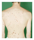
Dysplastic nevi usually become clinically apparent at puberty or adolescence, but true dysplastic nevi have been described in prepubertal children.1,4 The dysplastic nevi continue to appear throughout life. Their number may vary greatly, from one to hundreds. In patients with multiple lesions, there is marked heterogeneity among lesions, resulting in a highly characteristic clinical phenotype (Figure 2). Counts of dysplastic nevi are highly correlated with total nevus counts (increasing as the total count increases), independent of a personal or family history of melanoma.5
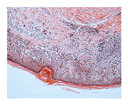
The term "dysplastic nevus syndrome" is used arbitrarily. Some clinicians use it to describe patients with only one atypical nevus,6 although the "classic" dysplastic nevus syndrome refers to patients who have the triad of 100 or more nevi, at least 1 nevus 8 mm or larger in diameter, and at least 1 nevus with clinically atypical features.7 The syndrome can occur sporadically or in a familial setting. To diagnose sporadic dysplastic nevus syndrome with certainty, clinical examination of first-degree relatives is necessary.8 Although there has been concern that nonspecialists might not reliably recognize the clinical phenotype, a recent study showed that identification of the phenotype with the use of a specific scoring system was a skill that could be easily learned by health care professionals who are not specialists.9 Pathological Diagnosis of Dysplastic Nevi The pathological diagnosis of a dysplastic nevus requires the identification of both specific cytologic and architectural abnormalities (Figure 3). Interobserver agreement in the pathological diagnosis of melanocytic dysplasia is hampered by the lack of universally accepted diagnostic criteria. In studies that use preset criteria for diagnosis, interobserver agreement is moderate to good.10,11,12 Reproducibility in grading atypia remains poor to moderate because of the nonuniform criteria used.11,12,13
Clinicopathological Correlations The presence or absence of atypical clinical features correlates, imperfectly, with the presence or absence of histologic dysplasia. In a study involving 101 patients with a history of sporadic melanoma in whom the most clinically atypical nevus was removed, the likelihood of histologic dysplasia ranged from 7 percent among lesions with no or only one clinically atypical feature (a diameter of 5 mm or more, an ill-defined or irregular border, or color variation) to 23 percent in cases with two atypical features and 62 percent in cases with three atypical features.14 Among nevi without histologic dysplasia, only 4 percent had all three clinically atypical features.14 In another study, roughly 30 percent of common (nondysplastic) nevi met the histologic criteria for dysplasia (architectural atypia, cytologic atypia, and a dermal host response), albeit minimally.15 In a population-based study, histologic findings of melanocytic dysplasia were associated with higher total nevus counts. Among subjects from whom biopsy specimens were obtained from the two most clinically atypical nevi, the histologic concordance between the biopsy results was higher than would be expected on the basis of chance alone, suggesting that melanocytic dysplasia is not randomly distributed but, rather, that some persons appear to have a predisposition to melanocytic dysplasia.16 Epidemiologic Characteristics The estimated prevalence of clinically atypical nevi ranges from 7 percent to 18 percent in population-based samples,17,18 and the prevalence of histologic melanocytic dysplasia is approximately 10 percent.19,20 The reported frequency of clinically atypical nevi among patients with a history of melanoma is higher, ranging from 34 percent to 59 percent.18,21,22,23,24 The variations in the estimates of prevalence probably reflect, at least in part, the use of different diagnostic criteria in different studies. However, differences among populations in sun exposure could also play a part.25 Dysplastic nevi also appear to be more prevalent in younger populations (those less than 30 to 40 years of age) than in older groups.5,8 Clinically atypical nevi may evolve from normal-appearing nevi or may be dysplastic from their first appearance.26 As dynamic lesions, dysplastic nevi can become progressively more or progressively less clinically atypical, but the majority either remain stable or regress over time.5,27 New dysplastic nevi may develop after the age of 30 years.5 The predilection of atypical nevi for intermittently sun-exposed areas (especially the trunk), their positive association with a history of painful sunburn (in which the pain lasts more than two days) or blistering sunburn, and the finding that persons with clinically atypical nevi often have sun-sensitive skin types suggest that the development of dysplastic nevi could relate to acute, intense sun exposure.3,18 Genetic factors also appear to be important; an autosomal dominant mode of inheritance has been reported in families with the dysplastic nevus�melanoma syndrome.8 Clinical Significance of Dysplastic Nevi The clinical importance of dysplastic nevi lies in their association with the risk of melanoma. This has been demonstrated in several cohort and case�control studies.17,18,19,20,21,22,23,24,27,28,29,30,31,32,33,34,35 In most of these studies, dysplastic nevi were defined according to clinical criteria, without histologic confirmation.21,22,23,24,27,28,30,31,32,33,34 In some studies, the presence of large nevi without other atypical features was also found to be associated with an increased risk of melanoma.22,24,35 The age-adjusted incidence of melanoma is approximately 15 times as high among patients with dysplastic nevi as among members of the general population (154 vs. 10 per 100,000 person-years, standardized to the population of Pennsylvania in 1985).31 The risk of melanoma in persons with dysplastic nevi increases with an increasing number of nevi and in the presence of a personal or family history of melanoma.29,30,31,32,34 There is a 100-fold increase in the incidence of melanoma in patients who have previously had melanoma, a 200-fold increase in those with at least two family members with melanoma, and a more than 1200-fold increase in those with both a personal and family history of melanoma.31 In persons with a family history of melanoma, the incidence of melanoma is highly concentrated in family members with dysplastic nevi, although melanoma may occur in those without this phenotype.28 The presence of dysplastic nevi is an independent risk factor for the development of multiple primary melanomas.28,36 Melanoma has been shown to occur both in and separate from preexisting nevi (common or dysplastic). In cohort studies of persons with dysplastic nevi, melanoma appears to arise in contiguity with dysplastic nevi in 44 to 80 percent of subjects,27,29,31,37 a rate much higher than that for melanoma in the general population.38,39 Currently, there are no published data on the extent to which the presence of histologic melanocytic dysplasia predicts melanoma independently of other risk factors for melanoma, such as clinically atypical nevi and the total number of nevi. Dysplastic nevi may be confused with melanoma, both clinically and histologically. In one study, lesions histologically diagnosed as dysplastic nevi by an expert panel were diagnosed by other pathologists as melanoma in 21 percent of the readings (in situ in 86 percent of the cases), and thin or in situ melanomas were misdiagnosed as dysplastic nevi in 12 percent of the readings.40 Strategies and Evidence Prophylactic Excision According to the available evidence, the clinical diagnosis of dysplastic nevi (or dysplastic nevus syndrome) does not need to be confirmed histologically.21,22,23,24,27,28,30,31,32,33,34 Despite the recognized association between dysplastic nevi and the risk of melanoma, the majority of dysplastic nevi will never progress to melanoma.26 The relatively low absolute risk that a single dysplastic nevus will develop into melanoma is indicated by the following calculations: in a population of 10 million inhabitants in which the incidence of melanoma is 10 per 100,000 inhabitants per year, about 1 million people (10 percent of the population) will have one or more dysplastic nevi (with a mean of two such nevi). On the assumption that 20 percent of melanomas develop in contiguity with a dysplastic nevus, this calculation implies that only 1 in 10,000 dysplastic nevi per year will progress to melanoma. These estimates underscore the argument that prophylactic excision of dysplastic nevi is unlikely to be cost effective and might, in addition, give patients a false sense of security, since an increased risk of melanoma may remain after these nevi have been removed. Some have advocated the excision of dysplastic nevi that are difficult to follow because of their location; however, data are lacking to support this approach.2 Follow-up of Patients with Dysplastic Nevi Melanoma of the skin offers the theoretical advantage of early detection by simple, noninvasive investigation (examination of the skin), and therefore regular dermatologic examinations are commonly recommended for persons with dysplastic nevi. In several studies, monitoring by trained dermatologists, with follow-up schedules ranging from once every three months to once a year, has been associated with high proportions of thin melanomas with a good prognosis,27,29,31,32,37 although those studies did not include contemporaneous control groups. Serial photographs have also been proposed as a method of documenting changes in existing lesions or the development of new lesions.29,31,32,41 In a historical, uncontrolled cohort study of patients with dysplastic nevi, 6 of 11 melanomas were excised because of change, relative to base line, in a preexisting nevus (in 5 cases) or development of a new lesion (in 1 case) on serial photographs; only 2 of the 11 lesions were diagnosed clinically as melanoma at the time of excision.31 Although close follow-up seems reasonable, it remains possible that close surveillance may preferentially detect slow-growing tumors with a good prognosis. In addition, it is uncertain whether the in situ melanomas (Clark level I) detected by such surveillance would have progressed to invasive melanoma if left in place. There are no prospective cohort studies comparing the survival of patients who are being closely monitored with those who are not. Proposed benefits of close surveillance include the opportunity to teach patients to identify suspicious lesions and possible improvements in the quality of life resulting from the reassurance of regular expert skin examinations; however, there are currently no data to support these possibilities. Prospective studies have demonstrated that the risk of melanoma in members of families affected by the dysplastic nevus�melanoma syndrome is considerable, with an estimated cumulative risk of 49 percent for persons 10 to 50 years of age and 82 percent by the age of 72 years27; the benefit of close surveillance is likely to be higher among such persons than among those with one or more dysplastic nevi but no family history of melanoma (which is the more frequent scenario). No studies have assessed the cost effectiveness of surveillance programs for dysplastic nevi. Self-Examination No randomized, controlled trials have assessed the value of self-examination of the skin. One study, which included 650 patients with melanoma, suggested a survival benefit in patients who had performed self-examination of the skin, as compared with those who had not, although the investigators emphasized that longer follow-up was needed to confirm a beneficial effect of self-examinations.42 In a survey of 816 patients with melanoma, the habit of skin self-examination was associated with thinner melanomas.43 Since self-examination is relatively simple and inexpensive, it is routinely recommended, despite the limited supporting data. Ocular Examination Patients with dysplastic nevi may have an increased risk of ocular melanoma.44 In a case�control study involving 211 patients with ocular melanoma, 16 percent had one or more dysplastic nevi, as compared with 7 percent of the controls (odds ratio, 24.1 [95 percent confidence interval, 4.8 to 119.8] among patients with four or more atypical nevi).44 The estimated cumulative lifetime risk of eye melanoma in patients with the dysplastic nevus phenotype is 1 in 200.44 A yearly ocular examination of patients with the dysplastic nevus syndrome has been recommended by some experts45 but not by others.46 Genetic Screening Germ-line CDKN2A mutations (on chromosome 9p21 ) have been found in 20 to 40 percent of families in which at least three first-degree relatives are affected by melanoma.47 These germ-line mutations were three times as likely to be found in family members affected by the dysplastic nevus syndrome as in relatives who were unaffected; however, the presence or absence of the syndrome was not perfectly correlated with gene-carrier status.48 Thus, a consortium recommended in a consensus statement that DNA testing not be performed in persons with the dysplastic nevus syndrome (familial or not) unless it is in the context of a defined research program, since the implications for management are currently unclear.47 Areas of Uncertainty Use of Sunscreens Total nevus counts have been found to be higher in children protected by sunscreens than in those protected by clothing,49 and a recent study50 indicated that the use of broad-spectrum sunscreens was effective in reducing the development of nevi in white children with freckles. However, data are lacking on the relation between the use of sunscreens and dysplastic nevi, and it is not known whether the use of sunscreens reduces the risk of melanoma.50,51 A real concern is that sunscreen use may lead to increased sun exposure, which could increase the risk of melanoma. Patients should thus be advised to avoid sun exposure during the hours of peak intensity of ultraviolet (UV) B radiation (noon to 4 p.m.) and to use physical protection, such as clothing, in addition to a broad-spectrum sunscreen (sun protection factor [SPF] 15 or higher) on uncovered areas. Dermoscopy Dermoscopy, or dermatoscopy (epiluminescence or incident-light microscopy), is a noninvasive technique in which oil immersion and optical magnification are used to make the epidermis translucent and to allow the visualization of structures not visible to the naked eye. The use of this technique has been proposed as a means of distinguishing melanocytic from nonmelanocytic pigmented lesions and of differentiating benign, suspicious, and malignant melanocytic lesions.52,53,54,55,56 Studies of images of skin lesions (including dysplastic nevi and early melanomas) extracted from image data bases have suggested that this technique improves the number of correct diagnoses over that achieved by clinical examination,53 but its influence on the rate of excision of benign lesions or other clinical outcomes has not been studied. Guidelines Table 1 summarizes recommendations for the management of dysplastic nevi according to two published sets of guidelines.57,58
Conclusions and Recommendations When encountering a patient with clinically dysplastic nevi, as in the case described in the vignette, the physician should take a detailed personal history, including information on any history of skin or other cancer, prior excisions of nevi, episodes of sunburn, and UV-radiation exposure (e.g., during childhood while residing in a sunny region) and should ask whether any family members have or have had melanoma or dysplastic nevi. All first-degree relatives should be encouraged to undergo a skin examination. During the complete examination of the skin at base line (which should include examination of the doubly protected areas and the scalp), the total number of nevi should be evaluated and the presence or absence of atypical nevi assessed. Although the role of dermoscopy remains controversial, we use this technique to evaluate lesions that look suspicious clinically, in particular to rule out the presence of nonmelanocytic pigmented lesions. A lesion should be excised if, after careful examination, the dermatologist believes that melanoma cannot be ruled out; in such cases, we excise the lesion with a 2-mm margin. Prophylactic excision is not warranted for lesions diagnosed clinically as dysplastic nevi, since the probability that a single lesion will develop into melanoma is very low and since excision does not clearly reduce the overall risk of melanoma. Patients should be educated about the risk of melanoma and should be advised to watch for alarming signs (changes in a mole or findings according to the "ABCD" rule for melanoma detection [asymmetry, border irregularity, color variation, or a diameter greater than 6 mm]). Avoidance of sun exposure during the hours of peak UV intensity (noon to 4 p.m.) and routine use of sun-protective clothing, sunglasses, and broad-spectrum sunscreens (SPF 15 or higher) should be routinely advised. Although data to guide follow-up are lacking, we follow patients with the use of serial, standardized (digital), total-body photographs and close-up photographs of prominent atypical nevi (with a ruler placed next to the nevus). We recommend reevaluation six months after the base-line examination and then once a year, or more frequent follow-up if there is a personal history of melanoma. Patients should be advised to request an earlier visit if they judge a newly appearing lesion to be suspicious or a change in an existing lesion to be suspicious, according to the information they have received.
From the Department of Dermatology, Gent University Hospital,
Gent, Belgium. Address reprint requests to Dr. Naeyaert at the Department of Dermatology, Gent University Hospital, De Pintelaan 185, 9000 Gent, Belgium, or at [email protected]. References
|