Basic
Research
NEJM
|

Multiple sclerosis is the most common nontraumatic disorder of the central nervous system in young adults. Twenty years after the onset of disease, 90 percent of cases will have entered a progressive phase.
Strong circumstantial evidence implicates an autoimmune mechanism in multiple sclerosis. The results of family studies illustrate the importance of genes in the familial aggregation of the disease. However, most monozygotic twins are discordant for the disease, and the markedly reduced risk of susceptible populations in subtropical regions demonstrates the substantial role of environmental effects.
As in many putatively autoimmune diseases, susceptibility to multiple sclerosis is governed in part by class I and II regions of the HLA genes, but other genes are also involved. Genome-wide scans of linkage in families with multiple sclerosis have yet to identify a novel susceptibility gene. Similarly, approaches involving non-HLA candidate genes have not shown any unambiguous associations. The slow pace of progress is common to studies of complex trait disorders and appears to result from the heterogeneity of disease pathogenesis. Approaches that could increase the pace are warranted.
Effector Candidates
In an effort to identify the processes involved in multiple sclerosis, Lock et al.1 used microarray technology (Figure 1) to scan 1080 genes to determine whether they were up-regulated or down-regulated in the lesions of four patients with multiple sclerosis. A total of 126 genes were expressed at twice the levels in the brains of at least three of these four patients, as compared with the brains of two control subjects, and 39 of these genes were increased in the brains of all four of the patients. The investigators also identified 151 genes that were down-regulated. Whitney et al.2 conducted a similar search in two lesions from the brain of a single patient with multiple sclerosis and found 62 differentially regulated genes.
|
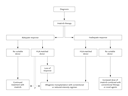
The large number of candidate genes identified in this way is daunting, and most of the differentially transcribed genes are not likely to be directly relevant to the pathogenesis of multiple sclerosis. It is to be expected that the expression of genes involved in inflammation, repair or remyelination, gliosis, apoptosis, cell signaling, and cell transport will be altered in the lesions of multiple sclerosis. Furthermore, there may be considerable time-dependent heterogeneity of expression profiles among lesions and perhaps within lesions. The selection of controls will be crucial, and many will be needed.
In a study of experimental autoimmune encephalomyelitis, an animal model of multiple sclerosis, Lock et al.1 selected two candidate genes that showed differences in expression depending on the type of lesion and tested them in mice with experimental autoimmune encephalomyelitis. Granulocyte colony-stimulating factor was overexpressed in acute, or active, plaques but not in chronic, or silent, plaques, whereas the Fc receptor was up-regulated in the chronic plaques and not increased in the acute plaques. Following these leads, Lock et al. found that both giving granulocyte colony-stimulating factor for six days before the induction of experimental autoimmune encephalomyelitis and inactivating the Fc receptor gene suppressed episodes of disease-induced inflammation in the mice. Using the same approaches for other candidate genes identified by microarray technology is a potentially powerful method of furthering research on multiple sclerosis. The resulting differences between acute and chronic lesions can help highlight the dynamic history of a lesion and identify appropriate therapeutic targets for a given stage of disease.
Candidate Susceptibility Genes
Microarrays could also be used to identify candidates for linkage and association studies and should be more discerning than identifying candidates purely on a speculative basis. This approach should reveal candidate genes that are up-regulated or down-regulated because of their direct involvement in causing multiple sclerosis or secondarily implicated in the primary event. But whether the candidate genes are part of a downstream process or the primary cause of multiple sclerosis, the knowledge gained from this approach will shed light on the overall pathogenesis of the disease.
Microarrays could also be used to study genomic regions thought to be linked to susceptibility. Microarray chips incorporating genes spanning such regions can be created. For example, there is evidence that loci in chromosomes 17 and 5 contribute somewhat to susceptibility. If all the genes from these regions were tested on a microarray chip, the resulting differences in expression could be used to identify a particular candidate gene. Candidate-gene approaches for complex diseases have not borne much fruit, but in the future there will be microarray chips for entire chromosomes and the entire genome. It cannot be assumed, however, that the expression of susceptibility genes would necessarily be altered in lesions at the time of sampling. Nevertheless, logical therapeutic design can concentrate on the effectors of tissue damage, the identification of which seems possible with the use of microarrays.
The Future
Characterization of plaques of different ages, from various locations within the central nervous system, and from various sources will provide the context in which to interpret results. The sample sizes should be as large as possible, and studies of serially obtained lesions would be highly desirable. Microarray data bases will also be invaluable. The microarray projects on multiple sclerosis have already collected vast amounts of readily available information, and data bases specific to multiple sclerosis or the central nervous system will provide a means of identifying the false positive findings that are likely to be generated. The data bases will also provide a framework for categorizing samples and results according to sex, HLA type, clinical course, and as shown by Lock et al.,1 the characteristics of the lesion. Future studies may involve a potentially huge systematic effort to sift through the many candidates identified by microarray technology, but since this approach is comprehensive, the chances of success are high.
David A. Dyment, M.Sc.
George C. Ebers, M.D.
University of Oxford
Oxford OX2 6HE, United Kingdom
References
- Lock C, Hermans G, Pedotti R, et al. Gene-microarray analysis of multiple sclerosis lesions yields new targets validated in autoimmune encephalomyelitis. Nat Med 2002;8:500-508. [ISI][Medline]
- Whitney LW, Becker KG, Tresser NJ, et al. Analysis of gene expression in multiple sclerosis lesions using cDNA microarrays. Ann Neurol 1999;46:425-428. [ISI][Medline]